How OCT Is Changing the Way Eye Doctors Detect Retinal Disease
79 Views
Just because you have blurry vision doesn't mean you need a new pair of glasses. Sometimes the real story is happening deep inside your eye, in the retina, where damage can build up quietly for years before you notice anything wrong. That delay is dangerous because most retinal diseases cause far more damage the longer they go undetected, and by the time symptoms show up, some of that vision loss cannot be reversed.
This is exactly the gap that optical coherence tomography (OCT) has closed. Any well-equipped eye hospital in Kokar Ranchi now uses this imaging technology to see inside the retina in a way that a basic eye exam simply cannot match, and any experienced retina specialist in Ranchi relies on it daily to catch problems early.
Keep reading to understand how this scan works, why it matters more than ever in 2026, and what it reveals that older diagnostic methods missed for decades.
What Exactly Is OCT and Why Does It Matter
Before getting into the technical details, it helps to understand what this scan actually does differently from a regular eye exam, and why eye doctors have grown so dependent on it over the past two decades.
The Basic Science Behind the Scan
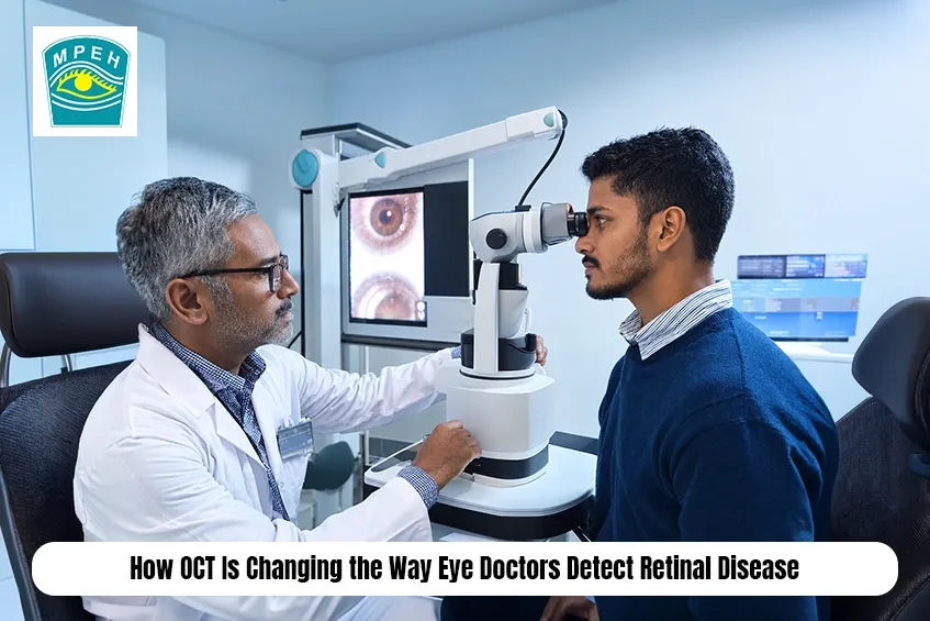
Optical coherence tomography works using light waves instead of X-rays or ultrasound. It sends a beam of light into the eye and measures how that light reflects off different retinal layers, then reconstructs those reflections into a detailed cross-sectional image. Think of it as in vivo histology, meaning doctors get a view of living retinal tissue that looks almost like a biopsy slide, except nothing is cut, injected, or removed. The scan takes just a few seconds per eye, involves no discomfort, and produces images with micron-level resolution, fine enough to detect retinal thickness changes as small as a few micrometres. That level of detail was simply unavailable to eye doctors relying only on a handheld ophthalmoscope or a slit lamp exam a few decades ago.
Why This Technology Became Essential in Modern Eye Care
A commonly searched question, " Why is OCT important for detecting eye disease?, deserves a direct answer. OCT reveals structural changes in the retina long before a patient notices any symptoms, which means conditions like macular degeneration, diabetic retinopathy, and glaucoma can be caught at a stage where treatment actually works best. Research tracking three decades of retinal OCT studies confirms that the technology's biggest research focus areas include retinal thickness measurement, therapies for macular degeneration and macular oedema, and the newer field of OCT angiography, showing just how central this scan has become to modern ophthalmology.
Structural Imaging Versus Vascular Imaging
Not all OCT scans do the same job, and this distinction confuses a lot of patients who assume one scan covers everything. Understanding the difference helps you know exactly what your doctor is checking during each visit.
What Structural OCT Actually Shows
Structural OCT captures the physical architecture of the retina, layer by layer. It measures thickness, detects fluid pockets, identifies swelling, and maps out the position of each retinal layer relative to the others. This is the scan doctors use to spot macular holes, macular cysts, vitreomacular traction, subretinal fluid, pigment epithelial detachment, and other structural abnormalities that a naked eye examination would likely miss entirely. It also plays a major role in evaluating choroidal neovascularisation, a condition where abnormal blood vessels grow beneath the retina and threaten central vision if left untreated.
What OCT Angiography Adds to the Picture
A frequently asked question worth answering here is: What is OCT angiography and how is it different from regular OCT? OCT angiography, often shortened to OCTA, goes a step further by mapping blood flow through the retinal and choroidal vasculature without requiring any dye injection. Traditional angiography needed fluorescein dye injected into a vein, which occasionally caused allergic reactions and added time to the appointment. OCTA skips that entirely, using motion contrast from moving blood cells to generate detailed vascular maps in seconds. This makes it especially valuable for detecting diabetic retinopathy, retinal vein occlusion, and abnormal vessel growth, giving doctors a non-invasive way to monitor blood flow changes over multiple visits without repeated dye exposure.
How OCT Detects Retinal Disease Earlier Than Ever
Early detection changes outcomes dramatically in retinal disease, and this section explains exactly how OCT manages to catch problems that older diagnostic methods routinely missed.
Catching Disease Before Symptoms Appear
Age-related macular degeneration (AMD) and diabetic macular oedema (DME) often progress silently in their early stages, with patients noticing no vision change until significant damage has already occurred. OCT changes that timeline by detecting microscopic fluid accumulation and structural distortion long before a patient reports symptoms. Deep learning research analysing thousands of OCT scans found that automated systems trained to identify retinal abnormality signs achieved accuracy rates ranging from 93% to 99% in distinguishing healthy retinas from diseased ones, a level of precision that continues to improve as datasets grow larger and imaging technology advances further into 2026 and beyond.
Monitoring Treatment Response Over Time
OCT does not stop being useful once a diagnosis is made. It becomes the primary tool for tracking how well a treatment is working. Doctors use repeated scans to measure whether retinal thickness is decreasing after anti-VEGF injections, whether macular oedema is resolving, and whether a previously identified area of fluid has dried up completely. This ongoing monitoring role is arguably just as important as the initial diagnosis, because it tells the treating doctor whether to continue the current treatment plan, adjust dosing intervals, or switch strategies altogether. Without this kind of objective, measurable feedback, treatment decisions would rely far more heavily on guesswork.
Swept Source OCT and Why It Represents the Next Generation
Not every OCT machine is built the same way, and the type of technology used can genuinely affect diagnostic accuracy. This section breaks down what makes newer scanning methods worth understanding.
The Technical Edge of Swept Source Technology
OCT technology has evolved through three main generations: time-domain, spectral-domain, and swept-source, each differing in scanning speed, image acquisition method, and resolution depth. Swept-source OCT represents the most advanced of these three, using a longer-wavelength light source that penetrates deeper into ocular tissue, including structures like the choroid that older machines struggled to visualise clearly. This deeper penetration matters because several retinal and choroidal conditions originate in tissue layers that shallower scans simply cannot reach with the same clarity.
Why Deeper Imaging Changes Clinical Decisions
A commonly searched question most people ask, "Is swept source OCT better than standard OCT?, comes up often among patients researching advanced eye care options. Swept source technology generally offers faster scan speeds and reduced motion artefact, meaning clearer images even in patients who struggle to hold perfectly still, which includes elderly patients and those with certain neurological conditions. It also allows for wider field imaging in a single scan, capturing more of the peripheral retina rather than focusing narrowly on just the central macula. For a retina specialist in Ranchi evaluating complex cases involving choroidal pathology or peripheral retinal disease, this expanded imaging depth often reveals findings that earlier generation scanners would have missed completely.
Conditions OCT Helps Diagnose and Manage
OCT is not a niche tool used for a handful of rare conditions. Its clinical applications span a wide range of common and sight-threatening retinal diseases, and this section covers the major categories doctors screen for regularly.
Common Retinal Conditions Identified Through OCT
Diabetic patients form one of the largest groups benefiting from routine OCT screening, since diabetic retinopathy and diabetic macular oedema remain leading causes of vision loss among working-age adults. OCT also plays a central role in diagnosing age-related macular degeneration, epiretinal membrane, retinal artery occlusion, and retinal vein occlusion, conditions that vary widely in cause but share the trait of being far easier to manage when caught early. Structural imaging also assists in evaluating vitreomacular interface disease, a category of conditions where the vitreous gel pulls abnormally on the retinal surface, sometimes requiring surgical intervention if left unaddressed.
Beyond Retinal Disease: OCT's Role in Glaucoma
Interestingly, OCT's usefulness extends beyond purely retinal conditions. It plays an equally important role in evaluating the optic nerve head, making it a standard tool for diagnosing and monitoring glaucoma alongside retinal disease. This dual capability means a single scanning session can assess both retinal health and optic nerve integrity, giving doctors a more complete picture of overall eye health in one visit rather than requiring separate specialised appointments for each concern.
Conclusion
Retinal disease used to be a waiting game, where doctors relied on visible symptoms and educated guesses until damage became obvious enough to diagnose confidently. OCT has rewritten that approach entirely by offering a noninvasive, fast, and remarkably precise way to visualise retinal architecture layer by layer. Structural OCT reveals swelling, fluid, and tissue distortion, while OCT angiography maps blood flow without any dye injection, and swept source technology now pushes imaging depth further than ever before. Together, these tools allow earlier diagnosis, more accurate monitoring, and genuinely better outcomes for patients dealing with diabetic eye disease, macular degeneration, and other sight-threatening conditions. Anyone experiencing blurred vision, distorted straight lines, or unexplained visual changes should not wait for symptoms to worsen. A proper OCT evaluation at a capable eye hospital in Kokar, Ranchi could catch a problem while it is still fully treatable, protecting vision that might otherwise be lost permanently.
Frequently Asked Questions (FAQs)
1. Is OCT scanning painful or invasive?
No, OCT is completely painless and noninvasive. It uses light waves, requires no injections, and takes only a few seconds per eye to complete.
2. How often should diabetic patients get an OCT scan?
Most eye specialists recommend annual OCT screening for diabetic patients, or more frequently if diabetic retinopathy or macular oedema has already been diagnosed.
3. Can OCT detect glaucoma as well as retinal disease?
Yes, OCT evaluates the optic nerve head structure, making it a standard tool for diagnosing and monitoring glaucoma alongside retinal conditions.
4. What is the difference between OCT and OCT angiography?
Standard OCT shows retinal structure and layers, while OCT angiography maps blood vessel flow without dye, revealing vascular abnormalities separately.
5. Does OCT replace the need for a regular eye exam?
No, OCT complements clinical examination. Doctors combine scan results with physical exams and patient history for a complete diagnosis.