Dental Photogrammetry vs. Intraoral Scanning: Which Is Better for Full-Arch Implants?
31 Views
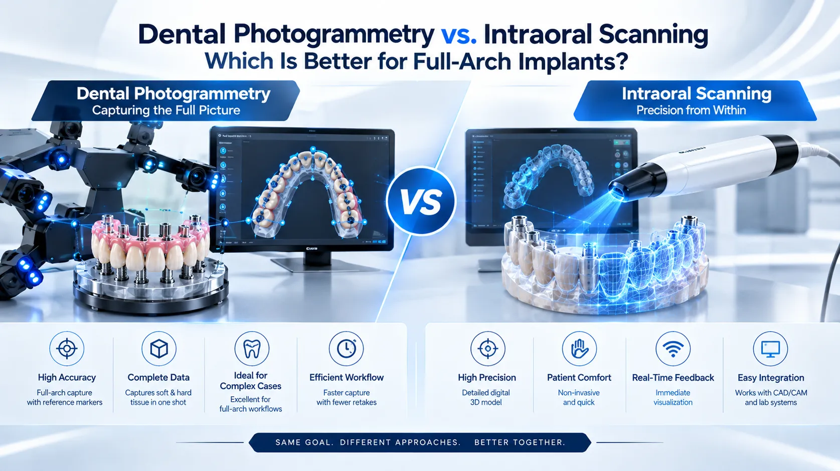
Quick Insight
For recording implant positions across a full arch, published research generally favors photogrammetry over intraoral scanning on accuracy, particularly angular accuracy. That does not make intraoral scanning obsolete. The two technologies capture different information, the evidence is still stronger in laboratory studies than in clinical ones, and neither removes the need to verify the result before the final prosthesis is made. Which one fits best depends on the case, the equipment available, and the workflow.
What Each Technology Actually Captures
The two methods are often framed as competitors, but they record different things.
|
Photogrammetry |
Intraoral Scanning |
|
|
Primary output |
Three-dimensional implant positions |
Surface anatomy: teeth, soft tissue, scan bodies |
|
How it works |
Calculates positions from overlapping images of coded markers or scan bodies |
Builds a surface model by optically capturing and stitching images |
|
Depends on intraoral landmarks |
No |
Yes |
|
Main strength |
Implant-position accuracy across the arch |
Soft-tissue and anatomy capture, widely available |
|
Main limitation |
Dedicated hardware and components |
Stitching error across long, edentulous spans |
In practice, many full-arch workflows use both: photogrammetry for implant position and intraoral scanning for tissue and opposing arch.
Why Intraoral Scanning Struggles on a Full Arch
ITI's clinical overview explains that the main limitation of intraoral scanning is how it reconstructs a model: best-fit algorithms stitch overlapping images by finding common reference features. On a fully edentulous arch there are few stable landmarks, and accuracy in full-arch implant cases, particularly in the mandible, remains debated. Photogrammetry avoids this by reconstructing positions from overlapping photographs rather than from intraoral anatomy.
Factors that tend to affect full-arch intraoral scans:
-
Long spans with no stable reference points
-
Soft-tissue movement and saliva
-
Scan body design and scanning technique
-
Mandibular versus maxillary arch
-
Operator scanning path
What the Accuracy Research Shows
A 2025 meta-analysis of 13 studies (3 in vivo and 10 in vitro) compared stereophotogrammetry with intraoral scanning for complete-arch implant impressions. Photogrammetry showed significantly greater trueness and precision overall, with reported angular trueness of 0.24° to 0.80° compared with 0.28° to 1.74° for intraoral scanning. The review noted that intraoral scanning reported angular deviation exceeding the 1° threshold associated with passive fit.
Important caveats from the same review:
-
Most included studies were in vitro, and the reviewers called for further clinical trials
-
Study methods varied widely, including reference standards and outcome measures
-
A few studies pointed the other way: one in vitro study found higher trueness but lower precision for intraoral scanning, another found higher accuracy with it, and one in vivo study showed comparable trueness
-
Until more clinical evidence exists, the review still recommends a rigid prototype try-in
What Clinical Evidence Adds
An in vivo JPD study compared a conventional intraoral scanner with an intraoral photogrammetry scanner in participants who each had four implants in an edentulous arch. Using a conventional splinted open-tray impression as the reference, the researchers analyzed 104 implant positions and reported that the photogrammetry scanner significantly improved both ease and accuracy of recording.
This study tested intraoral photogrammetry specifically, so it does not settle every comparison, but it adds in vivo support to the direction of the laboratory findings.
Types of Photogrammetry
-
Extraoral photogrammetry: a camera outside the mouth tracks coded scan bodies on the implants
-
Intraoral photogrammetry: the capability is built into an intraoral scanner, using coded markers on scan bodies
-
Navigated photogrammetry: emerging systems that link implant capture to navigation
Each has its own hardware, components, and workflow, so results from one system do not automatically apply to another.
How to Decide: Practical Factors
|
Factor |
Leans toward photogrammetry |
Leans toward intraoral scanning |
|
Arch and span |
Fully edentulous, long span, many implants |
Shorter span, stable landmarks |
|
Implant position accuracy needs |
High, especially angular |
Moderate, with a verification step |
|
Equipment already in the practice |
Photogrammetry system available |
Intraoral scanner only |
|
Workflow |
Willing to add components and a capture step |
Prefer a single-device workflow |
|
Soft-tissue capture |
Needs a companion scan |
Captured directly |
The same meta-analysis also cites a 2025 study concluding that both approaches can be clinically acceptable and that intraoral scanning offers a favorable balance of accuracy, efficiency, and cost. Cost and workflow are legitimate parts of the decision.
Where Fiducial Markers Fit
Photogrammetry depends on reference geometry the system can recognize. Fiducial marker systems supply that reference, and the ArchTracer fiducial marker system is one example designed for full-arch workflows. Marker design, count, and placement all affect how well any marker-based system performs.
Verification Still Matters With Either Method
Even the most accurate capture method does not guarantee a passive fit. Mixed results across studies, a reliance on in vitro data, and the cumulative nature of full-arch error all point to the same practice: confirm the transferred position before fabrication.
-
A verification jig or check scan compares the captured position against the clinical situation
-
A rigid prototype try-in confirms fit before the definitive prosthesis
-
Verification is workflow insurance regardless of which capture method was used
Manufacturers such as Digital Arches build verification and marker components for this stage of full-arch digital workflows.
Frequently Asked Questions
Is photogrammetry more accurate than intraoral scanning for full-arch implants? In published systematic review data, photogrammetry showed greater trueness and precision overall, but most evidence is in vitro and some studies found comparable or better results for intraoral scanning.
Does photogrammetry replace intraoral scanning? No. Photogrammetry records implant positions, while intraoral scanning captures surface anatomy, so many workflows use both.
Why is full-arch intraoral scanning harder than single-unit scanning? Scanners stitch images using shared reference features, and edentulous arches offer few stable landmarks over a long span.
Can intraoral scanning still be used for full-arch cases? Yes. Technique, scan body design, and verification all influence results, and it is commonly used with appropriate checks.
Do I still need a try-in if I use photogrammetry? The 2025 meta-analysis still recommends a rigid prototype try-in until more clinical trials are available.
Final Takeaway
On the evidence available, photogrammetry holds an accuracy advantage for capturing implant positions across a full arch, while intraoral scanning remains a practical, widely used tool whose results depend heavily on technique and case. The most defensible answer to "which is better" is that photogrammetry is generally more accurate for implant position, intraoral scanning is better for anatomy, and a verification step belongs in the workflow either way.
Related Reading
Expert Insights: Navigating the Complex World of Education & Learning
Welcome to our in-depth exploration of Education & Learning. In an era defin...
Why Education & Learning is Transforming the Global Industry Landscape
Welcome to our in-depth exploration of Education & Learning. In an era defin...